Patients suffering from oral ulcers often report that post-whitening enamel takes on a chalky hue, a phenomenon known as fluorosis aggravation. Before launching your next‐gen whitening device, B2B manufacturers and channel partners must understand how mucosal lesions interact with topical fluoride to avoid unintended contraindications and maintain clinical safety.
First, open lesions in the mucosa change fluoride dynamics:
Thus, ulcer management is pivotal to controlling fluoride uptake during whitening.
Next, consider the biochemical cascade:
This interplay underscores why ulcers are a red-flag contraindication.

Moreover, B2B partners must screen for factors that compound risk:
A thorough pre-treatment assessment avoids compounding fluorosis.
To mitigate fluorosis aggravation, integrate these design and chemistry controls:
Engineering both device and gel for targeted delivery preserves enamel integrity.
Clear usage protocols are essential:
Published contraindication lists in manuals ensure compliance at every touchpoint.
Finally, empower partners and safeguard reputation with:
A robust training and monitoring ecosystem turns potential risks into trust-building opportunities.
Conclusion
When oral ulcers and high-fluoride protocols collide, fluorosis aggravation becomes a real concern. B2B manufacturers must embed ulcer screening, pH-buffered gels, precise applicators, and strict contraindication protocols into their whitening solutions—supported by thorough channel training and surveillance—to guarantee both safety and efficacy. Contact us to co-develop clinically sound, user-friendly whitening systems that respect oral health complexities.
Oral care beauty trend :How Oral Care Became Beauty’s Sexiest New Category
5 Signs You Need to Replace Your Electric Toothbrush: Insights from an OEM Factory
.jpg)
Dental Clinic Toothbrush Bulk Supply | Reliable Oral Care Distributor
Why Is Toothbrush Quality Certification Non-Negotiable?
Indicator Failure Leading Periodontal Pocket Deepening – Undetected?
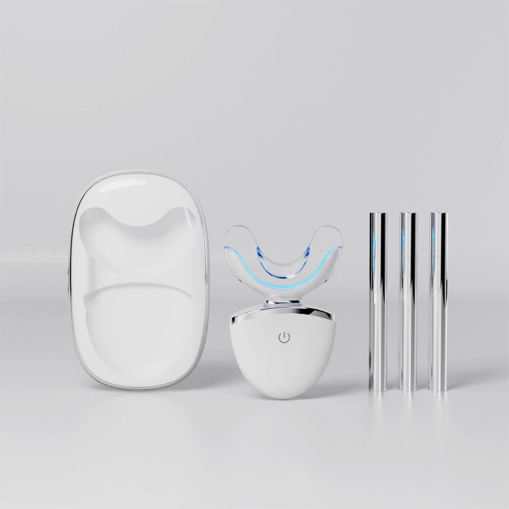
Home Use Teeth Whitening Device OEM for Global Oral Care Brands
How Can Servo Motor Customization Optimize Precision in Brush Head Injection Molding for B2B Toothbrush OEMs?
.jpg)
Proven Strategies to Unlock Premium Water Flosser Brands Positioning
Capitalizing on the LED Oral Care Boom: Essential Market Insights for Brands
Best Toothbrush for Doctors | Clinical Oral Care
Demineralization Zones with Jaw Fatigue – Silent Threat?
Can a Whitening Booster Solution Enhance Results from UV Whitening Technology?
.jpg)
sonic electric toothbrush Birmingham
Why is a Magnetic Drive Motor the ideal core for products requiring an IPX8 Waterproof Rating?
Split Bristles Linked to Enamel Cracks? 90% of Users Ignore This Danger!
Does Warranty Cover Motor Overheating in Electric Toothbrushes?

.jpg)
.jpg)
.jpg)
.jpg)
.jpg)
.jpg)
.jpg)